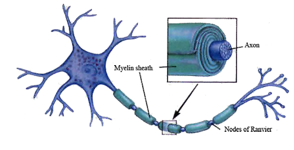
The mammalian nervous system is characterized by myelination, a fundamental biological process that protects the nerve processes of axons and facilitates the transmission of electrical pulses.
Damage to myelin is considered the major problem in MS. Currently, therapeutic interventions are focused on protecting myelin integrity and promoting myelin repair. These efforts need to be accompanied by an effective imaging tool that correlates the disease progression with the extent of myelination.
To date, magnetic resonance imaging (MRI) is the primary imaging technique to detect brain lesions in MS. However, conventional MRI cannot differentiate demyelinated lesions from other inflammatory lesions, and therefore cannot predict disease progression in MS.
To address this problem, these investigators prepared a gadolinium-based contrast agent, termed MIC that binds to myelin with high specificity.
MIC was useful for imaging of myelination in the rat brain.
MIC was found to distribute preferentially in highly myelinated regions and was able to detect regions of focally induced demyelination.
"This is an unmet need; we need imaging techniques to assess treatments that are targeting myelination. This will allow us to assess whether or not they are effective in early clinical trials."