A BRIEF BEGINNER'S GUIDE TO THE BRAIN AND MRI
The brain
The brain has three main parts: the cerebrum, the cerebellum and the brain stem. The cerebrum is the big part on the top which most people think of as the brain. The cerebellum is the smaller, roundish part that lies underneath the back of the cerebrum. The brain stem is the part that joins the brain to the spinal cord. It lies underneath the cerebrum, in front of the cerebellum and is at the top of the spinal cord.

The brain has three main types of “matter”, i.e. stuff. Gray matter is the stuff that does all the processing, encoding and storage – basically the “thinking”. White matter is the stuff that carries the signals between different parts of gray matter. If you imagine your phone connected to your computer via a USB cable, the phone and the computer are different parts of gray matter and the USB cable is the white matter allowing them to communicate with each other. Gray and white matter are made up of billions of neurons, or nerves: gray matter contains the main bodies of the nerves and white matter contains the tails of the nerves, called axons. Nerves that need to transmit fast signals have a myelin coating on their axons.

The third type of matter is CSF (cerebrospinal fluid). It bathes the brain, cushioning it and providing nourishment. There are reservoirs of CSF in the brain, the contents of which are continually refreshed by our bodies. These reservoirs are called ventricles. The biggest of these are called the lateral ventricles. If you slice the head from the eyes to the back of the skull and look down at it, you will see the lateral ventricles, one on either side of the line down the middle of the cerebrum, looking a bit like a butterfly.
Humans have evolved to have a lot of brain matter, so the only way to fit it all into the skull is to squeeze it together, like squishing up a piece of cloth to fit it into a tight space. This means that the cerebrum has lots of folds in it. The bits that go in are called sulci (a single sulci is called a sulcus) and the bits that go out are called gyri (a single gyri is called a gyrus) and they all have names. [Mammals that aren’t as intelligent as humans have smoother cerebrums, with fewer sulci and gyri, if any.]. Mice are a bit thick. You can see they have a massive cerebellum at the back (which controls balance and movement) and the two knobs at the front are the olfactory bulbs, because they are big on smell


The cerebrum has two halves, or hemispheres, which are approximate mirror images of each other. They are joined only by some tracts of white matter (like a bunch of USB cables instead of just one), so the two halves can work together. The main tract of white matter joining the two hemispheres is called the corpus callosum. It is in the middle of the cerebrum, next to the lateral ventricles. If you cut the corpus callosum, the left side and right side of the cerebrum cannot communicate properly. It is quite common for people with MS to have lesions on the corpus callosum. (A lesion is an area of damage or abnormality.)

Red bit is the corpus callosum, the highway between the halves of the brain
The cerebrum is split into four “lobes” (areas): frontal, parietal, temporal and occipital. Because the cerebrum has two halves, there are right and left frontal lobes, right and left parietal lobes, etc. The frontal lobe is more or less the front half of the cerebrum. It stops at the “central sulcus”. The parietal and temporal lobes lie behind the frontal lobe with the parietal lobe at the top of the head and the temporal lobes at the sides, more or less behind your ears. The occipital lobe is right at the back. The frontal lobe is important for personality, working memory, decision making, controlling inhibition, movement, etc. The parietal lobe is important for sensation, maths, music, humour, spatial tasks, etc. The temporal lobe is important for hearing, memory, language, emotions, object recognition, etc. The occipital lobe is dedicated to vision.

The outer layers of the cerebrum are made up of gray matter. Together, these layers are called the cortex or cerebral cortex. Anything to do with the cortex is referred to as cortical. (Anything to do with the cerebrum is called cerebral and anything to do with the cerebellum is called cerebellar.) White matter lying close to the cortex is called subcortical (or superficial). White matter lying deeper into the brain is called deep white matter.
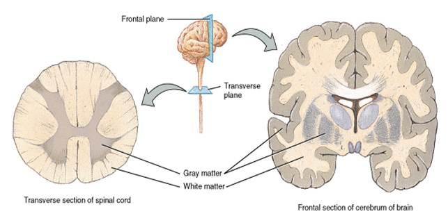
 There is also deep gray matter, found in the centre of the brain and the brain stem as well as white and gray matter in the spinal cord.
Amongst other things, the cerebellum is important for making all kinds of movement smooth, for balance and for learning new motor skills. The brain stem is important for many fundamental functions like sleeping, breathing and heart rate. Many of the “cranial nerves” come from the brain stem, controlling things like eye movements, sensation and movement of the face, mouth and tongue, and swallowing.

cranial nerves
In general, the left side of the cerebrum controls the right side of the body and vice versa. This is not the case in the cerebellum where the right side controls the right side of the body and the left, the left. All the nerves from the cerebrum and the cerebellum go through the brain stem to get to the spinal cord. The cerebral nerves cross in the brain stem so that the right side of the spinal cord controls the right side of the body and the left, the left. This means that a problem with the right side of the body might come from a left hemisphere cerebral lesion, a right hemisphere cerebellar lesion, the left or right side of the brain stem (depending on whether it is above or below where the nerves cross) or a lesion on the right hand side of the spinal cord.
The McDonald criteria for MS diagnosis stipulate that, to be diagnosed with relapsing remitting MS, patients need to have at least two attacks and at least one lesion in at least two of four specified areas of the central nervous system: three in the brain and one in the spinal cord.
The criteria for primary progressive MS are a gradual progression of symptoms (normally of at least a year’s duration) and two of the following: a positive lumbar puncture, at least two spinal cord lesions, at least one lesion in at least one of the three specified brain areas.
The three brain areas in the McDonald criteria are juxtacortical, periventricular and infratentorial. Juxtacortical means next to / into the cortex, i.e. a lesion that lies right at the border of the gray and white matter close to the outer edge of the cerebrum.
Periventricular means next to the ventricles. Lesions next to the lateral ventricles are very common in MS. They often form at right angles to the ventricles which, when looked at side on, look a bit like fingers poking up over a wall. These are called Dawson’s fingers and are a classic sign of MS. Infratentorial means the brain stem and cerebellum; the areas under the cerebrum.
There are some parts of the cortex that are named after what they do. The most obvious of these are the visual cortex, the motor cortex and the somatosensory cortex. The visual cortex is the cortex in the occipital lobe; the gray matter that makes sense of all the information that your eyes gather. The motor cortex is the last gyri of the frontal lobe, the precentral gyrus. It is in charge of movement. The somatosensory cortex is the first gyri of the parietal lobe, the postcentral gyrus. It is on the other side of the central sulcus to the motor cortex and it controls sensation in your body. If you put your hands loosely on both ears, your thumbs are pointing towards your visual cortex, your left and right ring fingers are pointing roughly towards your left and right motor cortices and your middle fingers are pointing roughly towards your somatosensory cortices.

There are some parts of the cortex that are named after what they do. The most obvious of these are the visual cortex, the motor cortex and the somatosensory cortex. The visual cortex is the cortex in the occipital lobe; the gray matter that makes sense of all the information that your eyes gather. The motor cortex is the last gyri of the frontal lobe, the precentral gyrus. It is in charge of movement. The somatosensory cortex is the first gyri of the parietal lobe, the postcentral gyrus. It is on the other side of the central sulcus to the motor cortex and it controls sensation in your body. If you put your hands loosely on both ears, your thumbs are pointing towards your visual cortex, your left and right ring fingers are pointing roughly towards your left and right motor cortices and your middle fingers are pointing roughly towards your somatosensory cortices.
It is rather complicated but different areas of the brain control different bits of the body.
Finally, a few terms that you might come across:
o Anterior: in front; towards the front of the brain
o Posterior: behind; towards the back of the brain
o Superior: above; towards the top of the brain
o Inferior: below; towards the spinal cord
o Horn/tip: refers to the ends of the lateral ventricles; the “wing tips” of the butterfly shape.
MRI: Magnetic Resonance Imaging
The MRI works by detecting the magnetic particles in atoms within cells and sending magnetic pulses at different rates and strengths of the electromagnetic pulse through the body. These are picked up by the Electromagnetic receiver. The magnetically charged particles in tissues get lined up with the magnetic pulse and then return (relax) to their positions once the magnetic field is turned off and this is detected by the machine.
There are long-strong pulses, short-strong pulses, and long- and short- weak pulses, and actually many more. The programmers use these different pulse/spin sequences to make the different tissue structures in the body stand out from each other. In 20+ years of studying the radiographers have discovered that different tissues (brain, bone, liver, blood, etc.) all show up best using different combinations of pulse techniques. They have also discovered that certain combinations of techniques show abnormalities like tumours or scars or whatever.
When you have an MRI scanning session, they run a number of different scans. The standard ones, that are run for pretty much all medical conditions, are called T1 and T2 (This depends on how they measure the relaxation of the magnetic particles). Scan types are constantly being developed, but the most commonly used additional scan type for MS investigations is FLAIR.
The way MRI works is that different types of matter give off different levels of energy when they are placed in a magnetic field. The computer slices the thing being scanned and collects the energy signal from one slice at a time. Because of some of the loud banging noises the scanner makes, it can also work out the signal from small cubes of each slice. These cubes are called voxels (short for volume pixels).

Each slice provides the information for one image and each voxel provides the information for one pixel in that image. The whole process uses some very advanced mathematics, but ultimately, the higher the overall signal from a voxel, the brighter the pixel is on the final image (and the lower the signal, the darker the pixel).
In a T1 scan, gray matter gives off a low signal and looks darker in the images than white matter which gives off a higher signal and looks pale gray. CSF meanwhile gives off the lowest signal and looks black.
Everything is reversed in a T2 scan: gray matter looks pale gray, white matter looks darker gray and CSF looks white. FLAIR is a clever adjustment of a T2 scan which suppresses the signal from CSF. This means that we end up with gray matter looking pale gray, white matter looking darker gray and CSF looking black.
FLAIR iss Fluid Attenuated Inversion Recovery. It is part of the T2 imaging, with a twist. Its purpose is to distinguish things that border on areas of fluid (such as CSF in the ventricles).
Different lesions give off different signals too. In general, a white matter MS lesion gives off a high signal in T2 and FLAIR scans, leading to it looking like a “white spot” against the white matter which looks relatively dark in these scan types.
T1 is usually rubbish for spotting MS lesions unless the lesion has caused the area to die, or “atrophy”, in which case it is called a “black hole” – because it is a black hole on a T1 image.
The terms “hyperintensity”, “hyperintense”, “high signal”, etc, all refer to the fact that somewhere is brighter / whiter than it should be as hyper = more. “Hypointensity”, “hypointense area”, etc, refers to somewhere that is darker than it should be as "hypo" is less. Exactly what relevance these have depends on their size, shape, location and the type of scan though.
Remember that it was mentioned CSF bathes the brain?
That means that all the sulci of the brain are full of CSF and so there is a lot of white on T2 images. Spotting a lesion in amongst lots of perfectly normal white stuff can be tricky, however it is really quite easy in FLAIR images – because there shouldn’t be very much white; all the CSF is black. (Please note that some small white spots can be perfectly normal – they are usually blood vessels.)
Sometimes, neuros ask for a scan with contrast. This is a T1 scan taken after the patient has been injected with a “contrast agent”. This is usually gadolinium which looks bright white on a T1 scan. The central nervous system, i.e. the brain and spinal cord, is protected by the blood brain barrier (bbb) which stops things that might harm it from getting in; gadolinium normally can’t get through the bbb.
In MS, cells from the immune system get through the Blood brain barrier and attack the myelin coating of nerves in that area, causing inflammation and damage: a lesion. While this is happening, the lesion is called “active”, “enhancing” or “contrast enhancing”. The gap the immune system has caused in the blood brain barrier allows gadolinium to get in. If there are no breaches in the blood brain barrier there should be no bright white signs of gadolinium inside the brain or spinal cord. If there are, these show where there are breaches, in other words, where the immune system is actively causing new damage.
Contrast is used for two main reasons: to show up very new lesions (typically lesions newer than about two to six weeks) because these can be difficult to see on normal MRI and to help show which lesions are active and which are not as this can be important for deciding on meds.
Newer types of scans, e.g. DWI (diffusion weighted imaging) which measures water flow, can be used to do this too so contrast is being used less often these days.
Some terms you might come across:
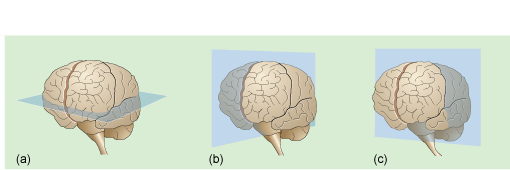
(a) Axial: images taken from front to back side to side, at right angles to the nose
(b) Sagittal: images taken from front to back, parallel to the nose
(c) Coronal: images taken from side to side, parallell to the nose and so that one slice could go through both ears
The 2 dimensional slice images taken through the brain are stacked to create a 3D image
o Artifact/artifact: a computer error, nothing to worry about
o 1.5T (and other numbers followed by a T): This is scanner strength, i.e. how strong the magnetic field is that the scanner produces. The T is short for Tesla, but has nothing to do with the T in T1 and T2 scans.
Most NHS scanners are 1.5T. There are a few 3T (and stronger) scanners that are much more powerful than 1.5T scanners. Do not assume that private MRI scanners (or scans) are better than NHS scanners (or scans) – they are mostly the same, but can be worse. There are also 7T very strong scanners usually run in some University Hospitals.
“Partial volume effects” are an important factor in how good a scan is and are particularly relevant if you are told your MRI is clear or you have no new lesions when you are having new symptoms. Remember that the computer slices up whatever’s being scanned and then splits the slices into little cubes, the signal from these determining how dark or light a pixel will be in the image? Well, the size of those cubes has a major effect. Imagine a voxel that only contains white matter.
In a T2 scan, that voxel will give a low signal and the resultant pixel will look dark gray. Now imagine a voxel that is mostly white matter, but also has a bit of a lesion in it. In a T2 scan, lesions give high signals, so the pixel will now look brighter than the last one. But by how much? If it’s a small voxel and the lesion provides a decent proportion of the signal, the pixel will be obviously brighter than its neighbour and a radiologist should spot it easily.
However, if it’s a big voxel, the extra signal from the lesion might only make the pixel a bit paler than its neighbour and make it very easy to miss. So, the moral of the story: it’s important to have thin slices and small voxels! 3mm slices are fine. Less than that is great, more than that and you are losing a lot of definition and increasing the chances of missing small lesions.By the time you get to 6mm and thicker, you could miss average and even bigger than average lesions too. (The average MS lesion is 7mm.)

The slice thickness is not usually written in reports or on images, but the number of images is the same as the number of slices so the greater the number of images in one scan, the thinner the slices. The best images generally come from thin slices with small voxels on a stronger scanner, but small voxels on a 1.5T scanner will be better than big voxels on a 3T scanner every time.
A01. Is this an oil rig, A02 a tank, A03 a car but I think we all see that A04 is a VW Beetle. So with a smaller the voxel size you get clearer pictures
So to recap
When you image these lesions with an MRI you can see different things, depending on the technique, the age (stage) of the lesion, the power of the MRI, and whether contrast is used. The first MRI image is done without contrast. Several different techniques are used in obtaining the images, including two called T1-weighted and T2-weighted. The first pass of the MRI will show old lesions that are big enough to be seen by the power of that MRI machine. WE KNOW that many lesions in MS are too small to be seen in some MRI's. As each new generation of MRI machines becomes available, it is more powerful and more able to show up areas of damage than the previous generation. If the newer, more powerful MRI more lesions will be seen
The "classic" old, scarred, mature MS lesion or plaque is somewhat oval in shape, will have well-defined borders and will appear in the white matter. The "classic" MS lesion will also have its long axis perpendicular to the ventricles (the large fluid-filled spaces) of the brain. Characteristic places (but not the only places) are peri-ventricular, juxtacortical, the corpus callosum, the cerebellum and the cervical spine. Also, important and often very symptomatic lesions are found in the brainstem and the thoracic spine. The spinal cord ends at the bottom of the thoracic spine, so there is no such thing as a lumbar spinal cord lesion. The scarred lesions will be evident as light, bright (hyperintense) areas on the T2 images. These are the classic MS lesions or "plaques." But, with just the regular MRI image one CANNOT say if it is old and dormant or if it has active inflammation in or around it.
arrows = MS plaques
Now the very old, damaged areas that have been reabsorbed will be seen as less dense (empty) spaces or "black holes" on the T1 images. If there are many of these empty areas, the brain will eventually contract and shrink around them. This will be depicted as a loss of brain volume. This is also known as brain atrophy. It is particularly seen in the progressive types of MS and after many years of disease. In brain atrophy there will be an increased space between the skull and the brain. The interior area of CSF will be larger. Also the deep folds in the brain (called sulci) will appear widened.
See the ventricles are much bigger in MS because parenchyma (paren key-ma) is being lost (BBF) Brain parenchymal fraction. This measures the space filled by the parenchyma (the tissue of the grey and white matter) as fraction of the whole brain voulume which contains fluid filled spaces. As the nerve tissue is lost the fluid filed spaces become bigger
The Need for Contrast
A newly active MS lesion may not be visible on a regular MRI because the area of nerves, though inflamed, is still pretty much intact and has normal brain density. On the regular MRI it will look like normal brain. Without contrast (gadolinium an magnetic compound) it won't show up and will likely be missed. When the next phase of MRI is done the contrast is injected into the bloodstream. Wherever the blood vessels are seen as more dilated than usual, bringing more blood to the area, (as in inflammation), the areas will "highlight" or "enhance." They show up as even brighter than the brain around them and brighter than an old, scarred lesion. So new lesions will appear as "enhancing," or "active." Also, older hyperintense lesions that have undergone a new attack right around them (also called reactivation) will show an even brighter enhancing rim or ring. The appearance of an "enhancing ring or rim" is especially characteristic of MS. When you compare the regular MRI to the contrast MRI you can see the increased brightness of this reactivated, old lesion. New lesions with active inflammation will typically show up for 2 to 6 weeks before they scar down and become "old" lesions.

Some radiologists call new lesion inflammation "active lesions." Others may refer to "new lesions " or "actively enhancing lesions." These all refer to the same thing. Also since some new lesions heal, the MRI's can be compared to old films where they did appear formerly, leading to the conclusion that they have disappeared. In addition, between different sets of MRI done after some time has passed, the radiologist can see an increase in old and in new activity. Right here it is important to remark that, in discussing MS, the word "active" is used in two different ways. If one is speaking about the appearance of lesions on the MRI, "active" simply means that the lesion has "active inflammation" in it. However, if we are discussing a person's disease and symptoms, an active lesion means one that is causing symptoms.
Another word that is used in two different ways in MRI reports is the word "new." If the radiologist is comparing the current MRI to an older one, a "new" lesion will be one that shows up now that didn't show up earlier. It has formed in the time period between the two MRIs. If the radiologist is describing lesions that show enhancement on an MRI, they might refer to these lesions as "new." The difference is in the context in which the word "new" is used.
In MS the Relapses and Remissions which are so typical of this disease occur because of the attacks on the nerves and then the body's ability to heal some or most of them. So we can often see the reflection of the disease process in the changes of the MRIs done over time.
The T2 technique is best for showing mature, dense, scarred lesions, where the oval lesions look brighter than the surrounding brain. This is where the term, "T2 hyperintensities" comes from. All T2 hyperintense lesions are not necessarily from MS. Other disorders like migraines, hypertension, small strokes and others can also cause them. On the other hand, the T1-weighted imaging technique is best for showing the old, reabsorbed "black holes" where the lesions once were.
To understand what the MRI is trying to see you need to have an idea on what is the disease pathology (Click HERE). The gadolinium enhancement has the best pathological correlate
http://multiple-sclerosis-research.blogspot.com/2012/04/education-espresso-pathology-grey.html
p.s. Thanks to V******* from down under for suggesting this
Want to Watch a video on this?
https://www.youtube.com/watch?v=xWFQ0eAGv24
https://www.youtube.com/watch?v=6X2HA0895HU
p.s. Thanks to V******* from down under for suggesting this
Want to Watch a video on this?
https://www.youtube.com/watch?v=xWFQ0eAGv24
https://www.youtube.com/watch?v=6X2HA0895HU


