There is a great need for the monitoring of microglial activation surrounding multiple sclerosis lesions as this is thought to be driving the widespread neuronal damage. Recently, 'second generation' positron emission tomography (PET) radioligands have been developed which can reveal the extent of microglial activation by quantifying the increased expression of the 18 kDa translocator (TSPO) protein. Here, we investigate whether PET imaging can be used to demonstrate the reduction in microglial activation surrounding a chronic focal multiple sclerosis (MS)-like lesion following treatment with fingolimod, an established MS therapy.
METHODS: Chronic focal experimental autoimmune encephalitis (EAE)-like lesions were induced in Lewis rats (n = 24) via stereotaxic intrastriatal injection of heat-killed bacillus Calmette-Guérin (BCG) and subsequent activation using an intradermal injection of BCG in complete Freund's adjuvant. This results in a delayed type hypersensitivity (DTH)-like EAE-lesion. The extent of neuroinflammation surrounding the lesion was measured using 18F-GE180 as a PET radioligand. The imaging was performed before and after treatment with fingolimod (0.3 mg/kg/day po, 28 days) or vehicle as a control. In addition to this, autoradiography and immunohistochemistry experiments were performed to verify the in vivo results.
RESULTS:The chronic DTH-EAE lesion led to increased ligand binding in the ipsilateral compared to contralateral hemisphere when PET imaging was performed with the TSPO-binding radioligand 18F-GE180. Treatment with fingolimod led to highly significant reduction in the binding potential, which could be demonstrated using both in vivo and ex vivo imaging (fingolimod vs. vehicle treatment, p<0.0001). The area of increased 18F-GE180 signal mapped closely to the area of activated microglial cells detected by immunohistochemistry.
CONCLUSION:PET-imaging, unlike magnetic resonance imaging (MRI), can be used to visualise the microglial activation surrounding a chronic DTH-EAE lesion. Importantly, treatment effect of fingolimod can be monitored in vivo by measuring the degree of microglial activation surrounding the chronic DTH-EAE lesion. This work gives promise for introduction of new outcome measures applicable in treatment studies of progressive MS.
This study suggests that we have a tool to measure hot microglia it is a short-activing radioactive tracer,in this can it is a 8 kDa translocator (TSPO) protein, another previously used is the benzodiazapine receptor. In this study the hot microglia were blocked by fingolimod. This will block the inflammatory penubra and blockinflammatory lesion formation,but what we need to block is the microglial inflammation in progressive disease. Can fingolimod do this in progressive MS, apparently not based on the recent failings in PPMS or are hot microglia a smokescreen, I somehow suspect not. Could this be useful in humans

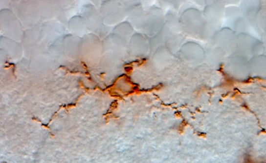
Image reveals higher levels of inflammation-associated translocator protein (orange/red) in the thalamus and other brain regions of chronic pain patients.
I guess so as images have now just been shown in humans in pain in the thalamus. Maybe this can be used to monitor progression in MS.