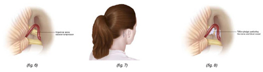
"How many of you have had MS-related trigeminal neuralgia? If you have you will know that it was of the most debilitating complications of having MS. The pain can be so severe it makes some people suicidal. The cause in MS has always thought to be due to a MS plaque, or lesion, in the brainstem where the trigeminal nerve enters the brainstem, i.e. the so called root entry zone. This study below suggests that it may be more than a MS plaque and that a blood vessel compressing the nerve may be contributing to the problem. We call this neurovascular compression and is what is thought to be the commonest cause of trigeminal neuralgia in people without MS."
"These findings suggests that in pwMS with intractable TN we will now have to look for neurovascular compression. Why? This form of TN may benefit from a surgical procedure that gently lifts the blood vessel off the nerve and puts a soft cushion between the vessel and the nerve. The cushion is believed to prevent the pulsations of the blood vessel activating the nerve fibres and causing the brain to perceive the signals as pain."
"This study is very timely, one of my patients presented last week with TN. I have ordered an MRI expecting to see a MS plaque in the pons; I wonder if the MRI will show a vascular loop pressing on the trigeminal nerve?"
Truini et al. A dual concurrent mechanism explains trigeminal neuralgia in patients with multiple sclerosis. Neurology. 2016 May 4. pii: 10.1212/WNL.0000000000002720.
OBJECTIVE: In this clinical and neuroimaging study, we sought information on the possible role of neurovascular compression in multiple sclerosis (MS)-related trigeminal neuralgia (TN).
METHODS: After screening 1,628 consecutive patients with MS, we enrolled 28 patients with definite unilateral MS-related TN. In these patients, we acquired dedicated 3T MRI scans, identified pontine demyelinating plaques, and, using highly specific diagnostic criteria, distinguished possible neurovascular compression.
RESULTS: MRI scans in most patients showed a demyelinating plaque in the pontine trigeminal root entry zone on the affected side. The frequency of the neurovascular compression and its association with the pontine demyelinating plaque were higher on the affected than on the unaffected side (54% vs 0%; p = 0.0001).
CONCLUSIONS: Our observation that in many patients with MS-related TN a pontine demyelinating plaque and neurovascular compression coexist should prompt neurologists to seek possible neurovascular compression in patients with MS-related TN.