If we can image demyelination, remyelination trials get a whole lot easier.
However you asked, Has this study in animals cracked the nut?
Updated
However you asked, Has this study in animals cracked the nut?
Updated
Brugarolas P, Sánchez-Rodríguez JE, Tsai HM, Basuli F, Cheng SH, Zhang X, Caprariello AV, Lacroix JJ, Freifelder R, Murali D, DeJesus O, Miller RH, Swenson RE, Chen CT, Herscovitch P, Reich DS, Bezanilla F, Popko B. Development of a PET radioligand for potassium channels to image CNS demyelination. Sci Rep. 2018; 8(1):607.
Central nervous system (CNS) demyelination represents the pathological hallmark of multiple sclerosis (MS) and contributes to other neurological conditions. Quantitative and specific imaging of demyelination would thus provide critical clinical insight. Here, we investigated the possibility of targeting axonal potassium channels to image demyelination by positron emission tomography (PET). These channels, which normally reside beneath the myelin sheath, become exposed upon demyelination and are the target of the MS drug, 4-aminopyridine (4-AP). We demonstrate using autoradiography that 4-AP has higher binding in non-myelinated and demyelinated versus well-myelinated CNS regions, and describe a fluorine-containing derivative, 3-F-4-AP, that has similar pharmacological properties and can be labeled with 18F for PET imaging. Additionally, we demonstrate that [18F]3-F-4-AP can be used to detect demyelination in rodents by PET. Further evaluation in Rhesus macaques shows higher binding in non-myelinated versus myelinated areas and excellent properties for brain imaging. Together, these data indicate that [18F]3-F-4-AP may be a valuable PET tracer for detecting CNS demyelination noninvasively.
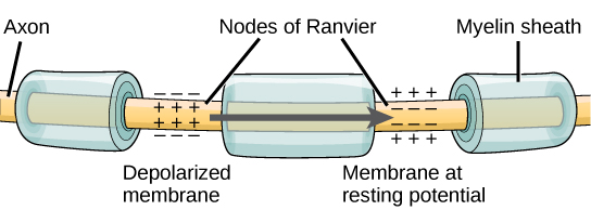

They investigated the possibility of targeting axonal potassium channels to image demyelination by positron emission tomography (PET). These channels, which normally reside beneath the myelin sheath, become exposed upon demyelination.
They use a variant of fampridine to target potassium channels and report that agent binds to non-myelinated areas better than myelinated areas.
The imaging agent in my mind however detects myelinated areas better than non-myelinated areas and binds all over the place. This because yellow is low binding, green is intermediate binding and red is high binding. Am I being a bit thick here and I am not interpreting this very well?
The argument in the paper is that there is low staining in the myelinated areas so it appears green and then when there is no myelin as occurs in the shivered the green areas become red

They made a variant of fampridine and tested it on a fly potassium channel which has similarity to Kv1.1, Kv1.2, Kv1.4 and Kv1.6 . It was excreted by the kidneys and liver but there was a window where it could detect areas covered with myelin.
However, there are a few problems I see. First it is detecting myelin by lower binding better than showing areas of active demyelination, which may require before and after images to truly show what was myelinated or look for the appearance of green.
The tracer shows fast entry into the brain and slow to moderate washout which is good. However, maybe another potential issue may be that voltage gate potassium channels are expressed all over the body and means the tracer with bind to potassium channels outside of the brain and it appears that there must be a lot in the kidneys as the signal does not disappear, which was seen in the monkeys (below). I will leave it for the imagers to determine how acceptable this is for humans.

PET of the Monkey you can see the line going in the arm and the kidneys lighting up.
The paper is open access so you can read it for yourself and make your own mind-up about how good the non-myelination/demyelination detection is.
Daniel ReichMonday, January 15, 2018 3:32:00 pm
said:
"Thanks for posting about our paper. The difference between this approach and prior myelin PET imaging methods is that [18F]3-F-4-AP is designed to highlight areas where there is less myelin, which in the white matter in MS would correspond to demyelinated areas. Other tracers bind to myelin itself, and therefore the images are diffusely bright throughout the white matter; finding a small area with loss of signal can be quite difficult. For all these approaches, interpreting the data will require knowing where the plaques are, and also knowing what is white matter and what is gray matter. A good MRI can tell you all of that, and the purpose of the PET scan would then be to help interpret what we see on the MRI. Testing whether this will work in practice awaits further experiments".

Daniel Reich (Click) – a neurologist and neuroradiologist – directs the Translational Neuroradiology Unit in National Institute for Neurological Disorders and Stroke. In his clinical practice, he cares for patients with multiple sclerosis and other neurological diseases, and he also leads several clinical studies focusing on multiple sclerosis. Research in his lab focuses on the use of advanced MRI techniques to understand the sources of disability in multiple sclerosis and on ways of adapting those approaches for research trials and patient care. He is particularly interested in harnessing noninvasive imaging modalities to dissect biological mechanisms of tissue damage.