Cerebellar abnormalities in early MS: the shredder may be quietly destroying your connections. #ClinicSpeak #MSResearch #MSBlog
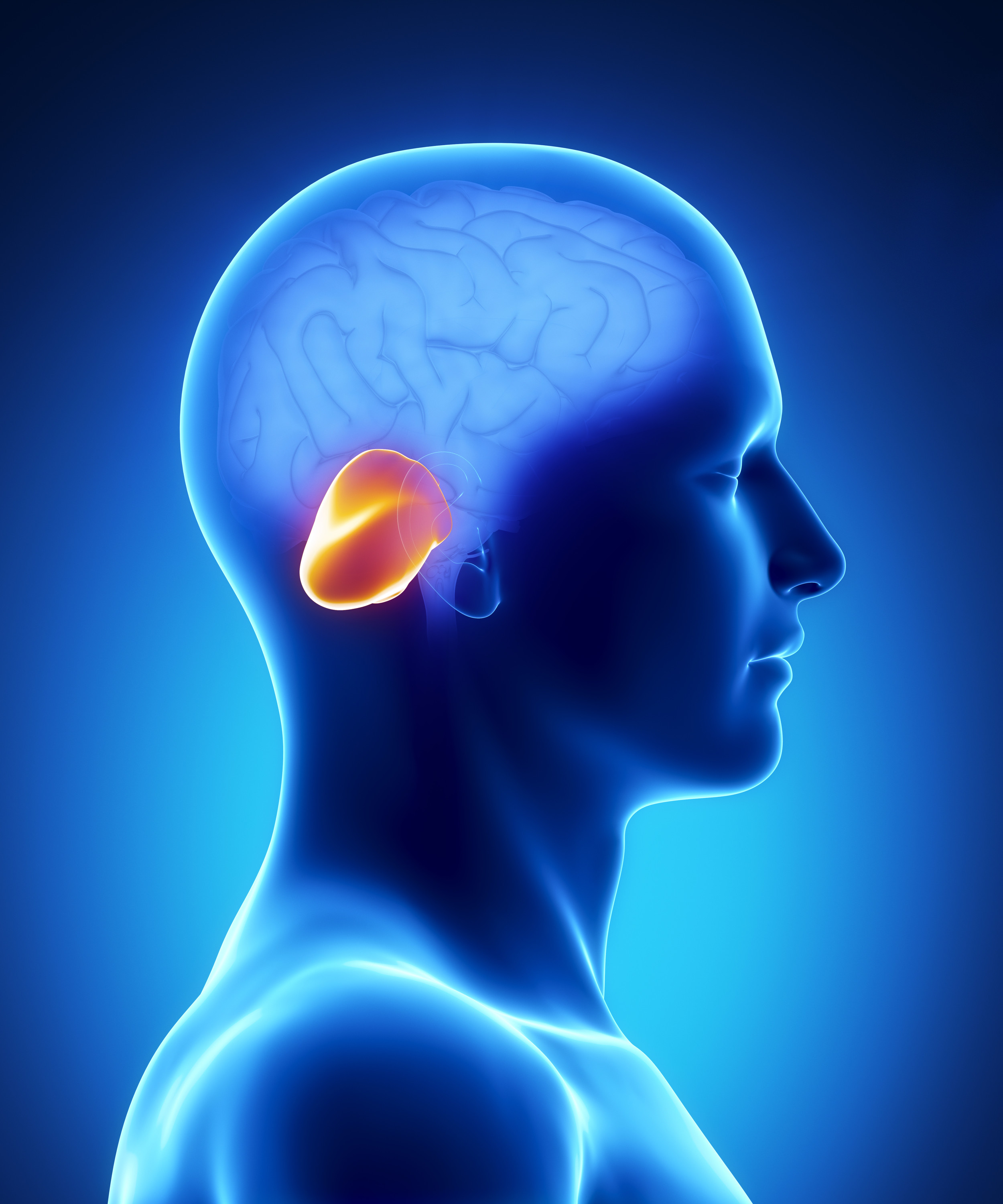
"The study below is very interesting in that it is applying new imaging technology to MS. It is using techniques to assess the connections of the so called cerebellar pathways in early MS. The cerebellum is the pathway that is mainly responsible for balance and coordination. The cerebellum is essential for the timing of movements. When I was at medical school I learnt a mnemonic for the symptoms and signs associated with cerebellar malfunction: VANISHDDD."
V = vertigo; dizziness with a feeling of rotation.
A = ataxia; unsteadiness of gait. We test for this by asking you to heel-toe walk on a straight line with your eyes open and the with your eyes closed.
N = nystagmus; jerking movements of the eyes, particularly when looking either left or right. Often the eyes jerk from when looking straight ahead; if you look closely there is a slow drift and a rapid correction. Neurologists call these movements square-wave jerks.
I = intention tremor; tremor of the fingers or hands when doing some; the tremor gets worse as you approach the target.
S = slurred speech; the speech sounds as if you have too much to drink. In fact alcohol is cerebellar toxin and a large number of the symptoms and signs of alcohol intoxication are due to cerebellar dysfunction.
H = hypotonia; this is when passive movements of the legs and arms suggest they are floppy.
D = dysmetria; difficulty judging distances. For example when you try and touch something you stop too early or you move beyond the object. If you have ever been asked to do the finger-nose test you point past the finger (past-pointing) or you miss your nose.
D = dysdiadochokinesia; this is a big word for clumsy movements. You may have been asked to do rapid alternating movements of the hands. If you can't do these and/or they are slow and clumsy we call this dysdiadochokinesia.
D = dysarthria; this is medical term for slurred speech.
Another sign should be cognitive impairment. Cognitive impairment is very common in MSers with severe cerebellar signs. In my experience it manifests as difficulty sequencing memories; cerebellar patients are very vague about when things happened and how they happened in relation to each other. Some of you may recognise these symptoms."
 |
| Cerebellum |
"What this study shows that in early MS, compared to controls, there are abnormalities in relation to the cerebellar connections. These abnormalities are found in early MS when there are no, or very few subtle signs of cerebellar dysfunction. What it is showing that MSers are able to compensate for the damage in their cerebellar pathways by using their reserve capacity or adaptations in other neuronal systems. These processes are called plasticity in neurospeak. Plasticity or the ability of the nervous system to cope with damage and is what keeps progressive MS at bay. Once the reserve capacity becomes exhausted then you manifest fixed neurological deficits that tend to get worse with time. This is another indication that we simply can't rely on the clinical examination to tell us that the brain and spinal cord are normal. What we need to use are methods, such as the MRI study below, to interrogate the functional reserve of the neuronal systems. It should be our therapeutic aim to protect and enhance this reserve to keep progressive disease at bay for as long as possible. The latter is a central theme of my asynchronous progressive MS hypothesis, i.e. there are multiple therapeutic windows to target even if once system has already exhausted its reserve capacity. For example, someone may have entered clinically apparent secondary progressive MS phase with weakness and difficulty walking in their legs, but still have very good upper limb and cognitive function. Should we simply write-off their upper limb function and cognition because their lower limb reserve capacity is exhausted? Of course not; this is why we have to design new trials for progressive MS that use outcome measures that focus on neuronal systems with reserve capacity."
"This post reminds me that we should be hearing about the headline results from the fingolimod PPMS trial very soon; I was told there would be a press release either in the last week of November or the first week of December. I wonder if my prediction of the results will be correct; this prediction is based on the asynchronous progressive MS and therapeutic lag hypotheses."

Romascano et al. Multicontrast connectometry: A new tool to assess cerebellum alterations in early relapsing‐remitting multiple sclerosis. Human Brain Mapping, 2014.
Background: Cerebellar pathology occurs in late multiple sclerosis (MS) but little is known about cerebellar changes during early disease stages. In this study, we propose a new multicontrast “connectometry” approach to assess the structural and functional integrity of cerebellar networks and connectivity in early MS.
Methods: We used diffusion spectrum and resting-state functional MRI (rs-fMRI) to establish the structural and functional cerebellar connectomes in 28 early relapsing-remitting MS patients and 16 healthy controls (HC). We performed multicontrast “connectometry” by quantifying multiple MRI parameters along the structural tracts (generalized fractional anisotropy-GFA, T1/T2 relaxation times and magnetization transfer ratio) and functional connectivity measures. Subsequently, we assessed multivariate differences in local connections and network properties between MS and HC subjects; finally, we correlated detected alterations with lesion load, disease duration, and clinical scores.
Results: In MS patients, a subset of structural connections showed quantitative MRI changes suggesting loss of axonal microstructure and integrity (increased T1 and decreased GFA, P < 0.05). These alterations highly correlated with motor, memory and attention in patients, but were independent of cerebellar lesion load and disease duration. Neither network organization nor rs-fMRI abnormalities were observed at this early stage.
Conclusion: Multicontrast cerebellar connectometry revealed subtle cerebellar alterations in MS patients, which were independent of conventional disease markers and highly correlated with patient function. Future work should assess the prognostic value of the observed damage.