The blood-brain barrier (BBB) provides a constant homeostatic brain environment that is essential for proper neural function. An unusually low rate of vesicular ( vesicle = blob of cell membrane) transport (transcytosis) has been identified as one of the two unique properties of CNS endothelial cells, relative to peripheral endothelial cells (the other is tight junctions which creates impermeable joins between cell), that maintain the restrictive quality of the BBB. However, it is not known how this low rate of transcytosis is achieved. Here we provide a mechanism whereby the regulation of CNS endothelial cell lipid composition specifically inhibits the caveolae-mediated transcytotic route readily used in the periphery. An unbiased lipidomic analysis reveals significant differences in endothelial cell lipid signatures from the CNS and periphery, which underlie a suppression of caveolae vesicle formation and trafficking in brain endothelial cells. Furthermore, lipids transported by Mfsd2a establish a unique lipid environment that inhibits caveolae vesicle formation in CNS endothelial cells to suppress transcytosis and ensure BBB integrity.


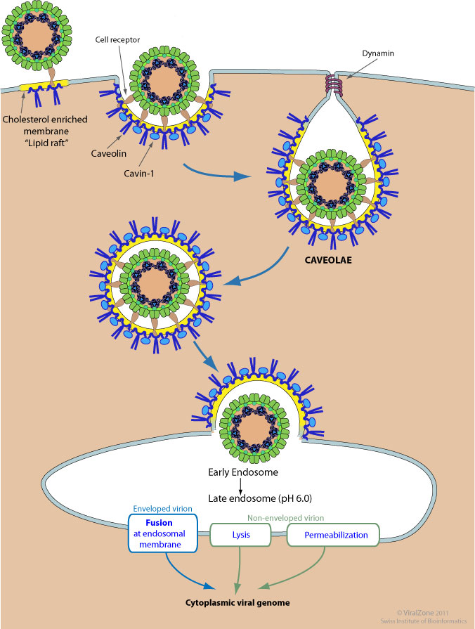
Caveolae (Latin for "little caves"), which are a special type of lipid raft, are small (50–100 nanometer) invaginations of the plasma membrane in many cell types, especially in endothelial cells.These flask-shaped structures are rich in proteins as well as lipids such as cholesterol and sphingolipids and have several functions in signal transduction.They are also believed to play a role in endocytosis.

They may be able to manipulate things to get treatments into the brain